MGD Innovations: Backed by research, powered by patients
Estimates describe over 30 million Americans having dry eye. That number exceeds 350 million worldwide.1
Meibomian Gland Dysfunction (MGD) is the most common cause of evaporative dry eye and has been the most challenging to effectively address clinically. Traditional therapies have failed to consistently provide effective results leading to ongoing suffering and frustration for patients and physicians alike.
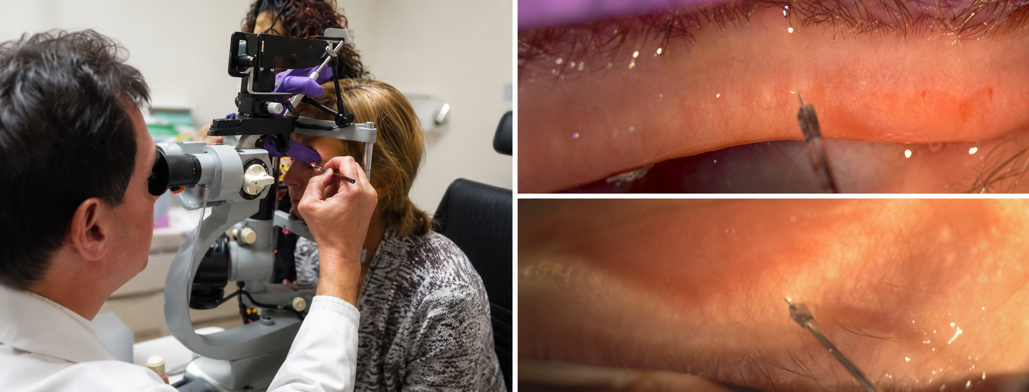
Meimbomian Gland Probing (MGP) was born out of necessity. While other therapies include drops (temporarily supplementing the tear film, reducing inflammation, or treating parasitic overgrowth), and devices that attempt to address the glands with heat and pressure from outside the eyelids, MGP was founded on extensive research and developed to address the root cause of MGD. MGP directly addresses the glands where this progressive disease hinders anatomical function.
Watch a short introduction to the Maskin Meibomian Gland Probing (MGP) Protocol.
Meibomian gland probing takes a different approach that aims to mechanically restore gland duct patency. Patients tell the story themselves… Below is a plot illustrating patient analog scoring of their own symptoms, starting at no (0%) improvement prior to therapy and maintenance of their symptom scores after probing. Improvements are demonstrated for patient groups with and without lid tenderness, where patients with lid tenderness are most likely to express a sense of immediate relief. Those without lid tenderness experience comparable long term benefits.
A summary of clinical publications is available in the links to the left.

Current procedures to clear glands of obstructions and promote oil secretion do not treat the underlying problem of periductal fibrosis (scar tissue) constraining Meibomian Glands and thereby overlook this long-term problem that progresses with the disease condition.
Common device-based procedures for dry eye, such as heat combined with pressure may reduce the presence of thickened meibum yet are not dilating the constrictions within glands. Releasing the fibrotic constriction, not just thickened meibum, is often necessary otherwise the gland remains blocked. MG probing not only releases thickened meibum and opens the duct, it releases the fibrotic constrictions around glands to target durable restored functionality.
Meibomian Gland Probing (MGP) is designed to clear glands of periductal fibroses allowing glands to again deliver essential meibum.
The plot below illustrates patient relief for those complaining of lid tenderness and evaluating lids with varying degrees of atrophy, measured using meibography.

MG Probing in Practice
Probing Meibomian Glands to return function and relieve dry eye symptoms is an essential component of a comprehensive treatment plan.
First, assess the health of the glands. Then, restore the integrity of the intraductal space through Meibomian Gland Probing. Finally, proceed with treatment for underlying and co-morbid factors of dry eye and MGD.
Evaporative dry eye requires restoring the tear film and meibomian gland dysfunction (MGD) may be coupled with aqueous tear deficiency (ATD). Such cases benefit from a combination therapy such as MG probing and punctal occlusion.
MEIBOMIAN GLAND EXAMINATION TO IDENTIFY ATROPHY:
Meibography can be an effective way to determine the number of functional glands and identify atrophied glands. This method does depend on consistency in setup, equipment, and lighting. Video meibography is recommended to prevent misleading artifacts in images.
MG Probing and duct tortuosity: Dr. Maskin has recorded MG probing with video meibography, illustrating how the Maskin probes follow and remain within the duct.
MASKIN® PROBING PROCEDURE: